


Summary
Physiologically accurate models of resistance blood vessels that are capable of vasoconstriction and vasodilation offer an excellent in vitro platform for testing drugs for vascular diseases involving smooth muscle cells. Vanderbilt researchers have developed methods to fabricate resistance blood vessels that make such a biomimetic platform feasible and could even be used to develop artificial organs.
Addressed Need
Improper functioning of resistance blood vessels plays a major role in vascular diseases like hypertension and vasculitis, as well as in diseases like Alzheimer’s and Parkinson’s that are associated with communication between vasculature and nearby neurons. Together, these disorders affect over 1 billion people worldwide and annually result in almost $1 trillion in direct and indirect costs in the US alone [1,2]. As new potential therapies for these diseases emerge, organ- on-a-chip models that can accurately simulate biological reality are crucial for streamlining drug development by minimizing the time and expense involved in live animal testing. Meanwhile, platforms to manufacture biologically accurate vasculature are critical to developing artificial organs that function properly and can address the chronic shortage of transplant organs.
Technology Description
Vanderbilt researchers have developed a technology that enables the fabrication of 3D models of resistance blood vessels with physiologically accurate stiffness, size, cell organization, and cell composition. Additionally, the mechanical stimulation and various coatings employed by this technology ensure the proper formation of the medial layer of helically aligned smooth muscle cell “actuators” necessary for the contractility characteristic of constricting and dilating resistance blood vessels.
Competitive Advantages
This method creates resistance blood vessels with far greater morphological and functional fidelity to biological reality than existing models. These vessels enable full in vitro resistance blood vessel constriction and dilation and allow for more extensive in vitro testing of vasculature-related drugs than is currently possible. With in vitro testing being faster and as much as 30 times less expensive than testing in animal models, this method allows for more efficient drug development. In addition, this technology also has potential in manufacturing in vivo tissue grafts and artificial organs.
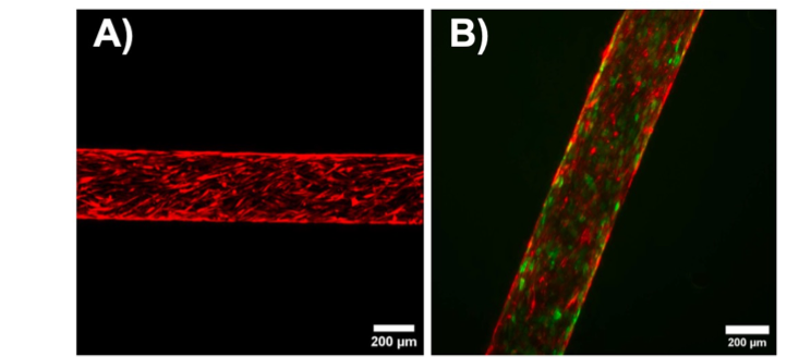
Microscopy images show that this technology properly aligns (A) smooth muscle cells and (B) smooth muscle and endothelial cells in arteriole-sized vessels, as indicated by cells being oriented 30-90 degrees from the vessel axis.
Stage of Development
Physiologically accurate resistance blood vessels with proper smooth muscle cell alignment have been developed and shown to contract and dilate in response to stimulants introduced via flow.
Intellectual Property Status
Patents: Patent application has been filed.